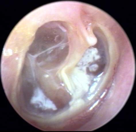
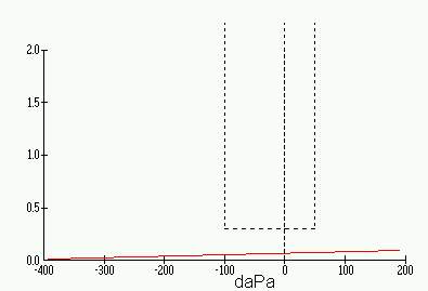
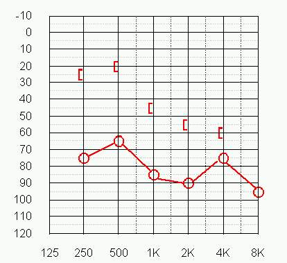
This AD TM image [inline below (18k)] was obtained from an 85 year old female patient with a lifelong history of chronic eustachian tube dysfunction and no prior hearing aid experience. The thin, atelectatic TM encases the malleus presenting with a prominent lateral process. There are centralized keratin deposits located anteriorly and postero-inferiorly with a light reflex visible on the promontory. The annulus is clearly demarcated around the severe TM injection (retraction). Absence of incus long process suggests resorption. A flat tympanogram(9k) with 1.7ml PVT is not inconsistent with the observed expansion of V3 into V4. AD pure tone air and bone conduction thresholds(29k) reveal a severe mixed loss with an 85 dB SRT and a 32% maximum word recognition score. Pure tone thresholds averaged 53 dB with 96% word recognition in the contralateral ear.