VO Case Presentations:
HYPERAEMIC SOFT TISSUE MASS IN POST/SUP EAM
Kevin Pons, M.S., FAAA
Yankton Medical Clinic; Yankton, SD
(Posted May 1, 2001)
|
The patient is a 42 year old female presented on video otoscopic examination with a large reddish (hyperaemic) mass arising from the epidermis of the posterior superior quadrant of the external auditory meatus. The lesion was broad based with an irregular surface. Computerized tomography (CT) verified a soft tissue mass arising from the posterior and superior external auditory canal wall, extending medially toward, but not beyond, the tympanic membrane. Audiometry and aural acoustic immittance were within normal limits.
The patient was moving out of town and was medically advised to seek oto-surgical consultation with extirpation and histological verification of the lesion at her new location.
ADDENDUM: The patient reported back that the lesion was succesfully removed surgically and histologically verified as a benign mixoid neurofibroma.

Figure 1 shows a large, hyperaemic mass in the EAM.
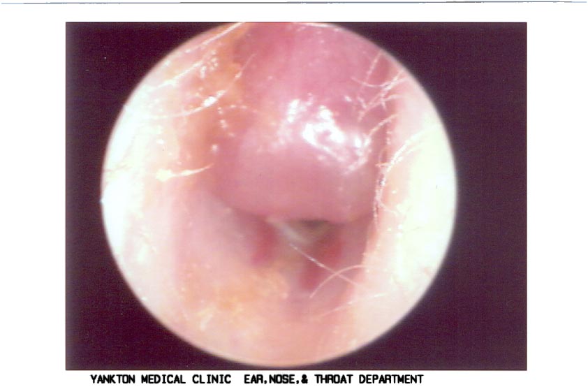
Figure 2 shows an alternative view of the above.
Case contributed by Kevin Pons, M.S., FAAA; Yankton Medical Clinic; Yankton, SD
posted May 1, 2001
|
|