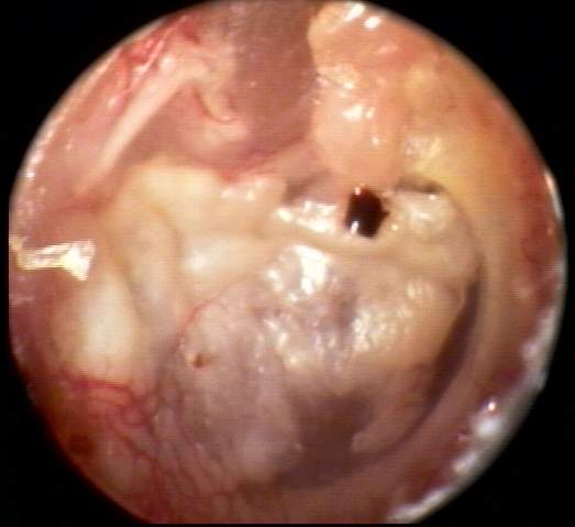
This AD TM image [inline below (20k)] was obtained from an 87 year old female patient presenting with symptoms of senile dementia and a six year history of hearing aid use on this ear.The instrument was recently lost. Extensive history of middle ear disease is assumed but could not be verified. The otic condition was diagnosed by the consulting ENT as tympanosclerosis with extensive, hyalinized collagen deposits throughout the TM. Tympanometry(17k) belies the abnormality. Acoustic reflexes were absent with probe AD. Unmasked pure tone air and bone conduction thresholds(31k) were obtained with difficulty. Masked results proved invalid. The presumptive air-bone gap is not inconsistent with the observed condition.