CASE STUDY: Crus Helicis Modification; Arthritic Digital ContracturesRoy F. Sullivan, Ph.D. (posted 11/02/1997 revised 03/09/1999) This 79 year old female patient with profound arthritic distortions of both arm joints and hands (inline image A below; 23k) was seen, after a two and one half year interval - overly long due to poor health- for a periodic follow-up evaluation. She had been fitted binaurally more than five years previously with CLASS III (unvented) full concha hearing aids. The right aid had been recently lost in a hospital stay.  The left aid, because of the anterior pressure caused by the patient's accomodative strategy for double height (2W) gain wheel adjustment (inline image B above)), was now found to be causing a contact ulceration of the crus helicis. (video oto-macroscopic inline image below, 25k). The dotted line showed the original anterior margin of the aid in the area of the crus. That area of the shell and faceplate was ground back with a hand motor tool and the resulting dehiscence in the shell repaired with monomer / polymer methyl methacrylate. 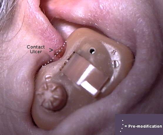 Because of a significant regression in hearing acuity, an impression was taken for a replacement left instrument. The patient declined a replacement of the lost right aid, having effectively lost the function of her right arm and hand. A Polaroid reproduction of the left crural area, shown above, was transmitted to the hearing aid laboratory with specific instructions concerning the fit. The new instrument is shown in situ, (inline image below 25k) ten days later. The contact ulceration had fully resolved.  Video oto-macroscopy (VOM) is invaluable in communicating the need for morphological changes in hearing aid design to the manufacturing laboratory.
|