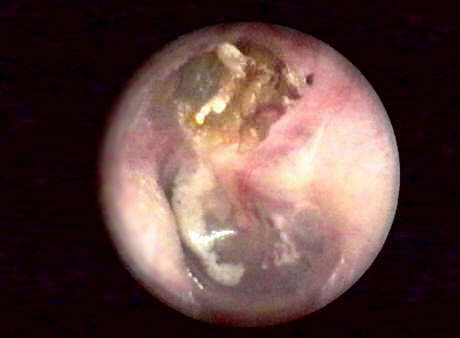
This 20 year old male patient presented with a unilateral 35 dB conductive hearing loss overlay. Video otoscopic examination revealed yellowish keratin debris in Shrapnell's area (pars flaccida) of the AS tympanic membrane, surrounded by "attic cerumen" often a characteristic sign of attic cholesteatoma. Keratin plaque is noted in the posterior inferior quadrant.
A CT scan is generally considered to be superior to MRI for the detection of choesteatoma-induced bone erosion. The site of lesion is noted in the axial view presented below. Special thanks to Thomas Lange, M.D. of Radiologische Gemeinschaftpraxis, Kiel, De. for most knowledgeably correcting my earlier image labeling errors.
