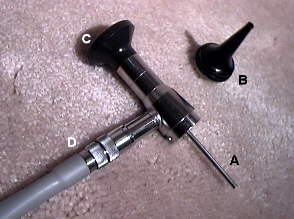
Fig. 4 shows an oto-endoscope or rod otoscope designed for ocular, rather than video, viewing at a fixed focal length.
Fig. 4. Fixed focus rod otoscope designed for ocular viewing. (A) endoscopic rod; (B) twist lock otic speculum; (C) ocular eyepiece; (D) fiber optic light bundle.
The stainless steel rod casing (Fig. 5) is typically 30 - 40 mm long containing a central solid rod lens which transmits the otic image and a circumferential fiber optic bundle which transmits the source light. The narrow diameter of the rigid rod, less than 3mm, facilitates an intimate examination of the TM and EAC. This differs from flexible medical endoscopes in which both image and source light are transmitted via concentric flexible fiber optic bundles.
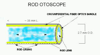
Fig. 5. Schematic diagram of the oto-endoscopic rod.
Otoscopic fields of view may vary, among manufacturers, from 10 degrees for some standard non-video otoscopes through 80 degrees for commercially available VO systems. With otic speculum in place, the tip diameter ranges from 4.0 - 5.0 mm, facilitating a full screen, circumferential image of the tympanic membrane. Fig. 6 shows a comparison of luminance levels, image size and spherical aberration among three different model VOE heads using the same light source and distance from the 1 mm calibration rulings.
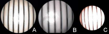
Fig. 6. Variability (A-C) in luminance levels, image size and spherical aberration among three different video rod endoscopes.
Fig. 7 identifies the features of a representative clinical video oto-endoscope. The endoscopic rod (A) is reinforced (B) to facilitate use of a disposable cerumen wax loop (D) as well as the autoclavable otic speculum (C). The Siegle bulb (E) is particularly important for visual verification of TM mobility, differentiating monomeric scars from perforations and for clearing condensations from the distal rod lens. The change from room (~68oF) to body (98.6oF) temperature at higher humidity levels often causes the VO lens to fog. Application of the bulb quickly restores a clear field of view. The fine focus (F) can be changed easily while the VO head is in situ. A quick release ring (G) permits rapid interchange between the standard VOE and the VOM lens systems.
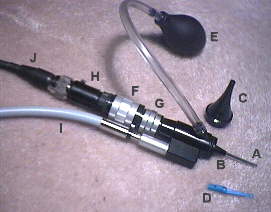
Fig. 7. Enhanced video otoscope: (A, B, I) Video oto-endoscopy (VOE) head; (A) endoscopic rod; (B) rod brace for cerumen curettage; (C) autoclavable otic speculum; (D) disposable cerumen loop; (E) Politzer bulb; (F) in situ focus knob; (G) quick release for video oto-macroscopy (VOM) lens; (H) 1/2" 2.5 lux S-video camera; (I) fiber optics source light cable; (J) camera cable.
Roy F. Sullivan, Ph.D.